flow cytometry results for lymphoma
Flow cytometric leukemia and lymphoma analysis may aid in identifying the tumor lineage for diagnostic and prognostic purposes. 88184 - 1st Marker.

Pin On Clinical Medicine Practical
Flow cytometry FCM has been used for rapid and sensitive diagnosis of various hematologic malignancies 34.

. WBC1700uL Hb89 gdL Plt168000uL Differential count. Case 1 have expression of B cellassociated antigens CD19 CD20 CD22 and CD79a by flow cytometry. Flow cytometry has become an important tool in the diagnosis of mature lymphoid neoplasms and the determination of prognosis in selected cases.
88185 - Each Remaining Marker. They also expressed the CD38 CD123 CD58 CD81 and HLA-DR. However flow cytometry results usually make certain lymphoma entities extremely likely and others very unlikely.
Phenotypic assessment of lymphoid cells can be done with flow cytometry. 88187 88188 88189 - Professional. Examples Of Cd200 Expression In Mantle Cell Lymphoma By Flow Cytometry Download Scientific Diagram.
FCM analysis has also been demonstrated as an excellent diagnostic tool for detecting leukemia and lymphoma cells in various body fluids 5 6. Flow cytometric immunophenotyping is useful in diagnosing lymphoma. Immunophenotyping Flow Cytometry for Hematolymphoid Neoplasia.
Results may be delayed or the sample rejected if pertinent andor required information is conflicting or missing. Two-color analysis primarily for surface markers is currently the standard method for flow cytometry measurements in routine diagnostic studies of leukemia and lymphoma. Three samples that came from patients who had morphologic evidence of malignant disease on biopsy two Hodgkins disease and one large cell lymphoma had flow cytometry results that were interpreted as normal.
11 lymphs including hematogones Cytogenetics. This is especially true if initial testing showed an increased number of lymphocytes abnormal cell counts or the presence of immature blood cells. These can be stratified as large and small lymphocytes CD45 positive.
Several recent studies that used either immunohistochemistry or molecular expression array profiling have demonstrated that specific patterns of the inflammatory milieu or antigen expression in HRS cells themselves correlate. Results from the flow cytometry show the detected CD numbers which doctors use to compare to regular and irregular cells allowing them to form a diagnosis. See Flow Cytometry Report from Aspirus Reference Laboratory.
Flow Cytometry Results Of Suspected Central Nervous System Download Scientific Diagram Anti Brdu Antibody Clone Bu20a Bio Rad Analysis Expressions Map Screenshot. Flow cytometry is rapid and appears to be virtually diagnostic of non-Hodgkins lymphoma when a majority of cells are B cells with an abnormal kappalambda ratio. This flow cytometry test is used to diagnose leukemia or lymphoma.
The advantages of flow cytometry are based largely on its ability to analyse rapidly and simultaneously multiple cell properties in a quantitative manner. Lineage identification can provide a confirmatory diagnosis or differential diagnosis prognosis and treatment options. The official flow cytometry labo- ratory report is most commonly an individual-lab-generated paper report form.
A discussion of the potential. The gating dot plot below identifies a predominant CD45 bright FS small used cells. Burkitts lymphoma BL is a cancer of the lymphatic system in particular B lymphocytes.
These cells were in the subsequent anlysis. Not always strictly speaking not very often. Case 1 lack the expression of CD10 CD15 NG2 CD3 cCD3 MPO CD13 CD33 and CD7.
It is used to detect abnormal hematolymphoid populations determine what cell surface markers they express and integrate immunophenotypic findings with morphologic and available clinical and. 5 segs 52 lymphocytes 32 monocytes 9 eosinophils. 47XX8t922q34q112146XX19 t922 translocation in 1 of 200 cells analyzed.
It is a highly aggressive lymphoma that is usually found in extranodal sites or presenting as an acute leukemia. Leukemia and lymphoma are both. This test generates a hematopathology report with a diagnosis and interpretation of findings.
Leukemias and lymphomas are caused by an abnormal white blood cell that begins to divide uncontrollably making numerous copies of itself clones. Briefly the cells are individually passed in front of a laser where they are further sorted and characterized. Leukemia and lymphoma analysis by flow cytometry aids in identifying the tumor lineage which in most cases is identified as T cell B cell or myeloid.
Flow cytometry to be discussed in detail in another Morning Report is a powerful technique that allows for phenotyping of unfixed cells in fluid. Results are reported by Aspirus Reference Laboratory in 2-4 working days. These results will explain if any abnormal cells are present and what types of cells they are as a part of your diagnosis.
When using fresh tissue for flow cytometric immunophenotyping the predominant populations are lymphoid. Flow cytometry immunophenotyping may be useful in helping to diagnose classify treat and determine prognosis of these blood cell cancers. Therefore flow cytometry is an important integral part of lymphoma diagnosis even in cases where it cannot give a definitive diagnosis.
In most cases the lineage can be identified as T-cell B-cell or myeloid and a. We hypothesize that flow cytometry could also be successfully applied to prognostication of clinical outcome of Hodgkin lymphoma. After review of the clinical history and morphology a panel of markers is selected for each case by a board-certified hematopathologist.
Click here for instructions on how to download the free FCS Express Reader to view and manipulate the sample cases. This test is usually done after atypical results are seen on a complete blood count or white blood cell WBC differential. Flow cytometry is generally used as follow up testing after a complete blood count CBC or white blood cells scan WBC.

Flow Cytometric Presentation Of A Large B Cell Lymphoma A Forward Download Scientific Diagram

Use Of Flow Cytometry In The Phenotypic Diagnosis Of Hodgkin S Lymphoma Grewal 2019 Cytometry Part B Clinical Cytometry Wiley Online Library

Flow Cytometry Results Of Suspected Central Nervous System Download Scientific Diagram

Demystifying The Diagnosis And Classification Of Lymphoma A Guide To The Hematopathologist S Galaxy Mdedge Hematology And Oncolo Oncology Diagnosis Lymphoma

T Cell Marker Detections In B Cell Lymphomas Other Than Cll Sll And Download Scientific Diagram

Use Of Flow Cytometry In The Phenotypic Diagnosis Of Hodgkin S Lymphoma Grewal 2019 Cytometry Part B Clinical Cytometry Wiley Online Library

Immunophenotype By Flow Cytometry Of The Peripheral Blood Showing Download Scientific Diagram

Examples Of Cd200 Expression In Mantle Cell Lymphoma By Flow Cytometry Download Scientific Diagram
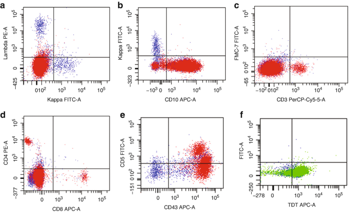
Flow Cytometry Of Mature And Immature T Cell Lymphoma Springerlink
Intra Tumour Heterogeneity Of Diffuse Large B Cell Lymphoma Involves The Induction Of Diversified Stroma Tumour Interfaces Ebiomedicine

Flow Cytometric Immunophenotyping Performed On The Same Plasmablastic Download Scientific Diagram
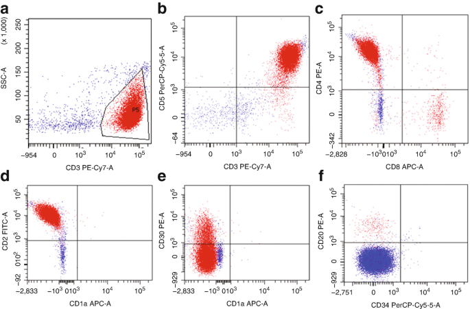
Flow Cytometry Of Mature And Immature T Cell Lymphoma Springerlink

Targeting The Tumor Microenvironment Of Leukemia And Lymphoma Trends In Cancer
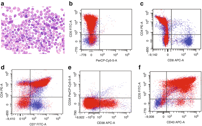
Flow Cytometry Of Mature And Immature T Cell Lymphoma Springerlink

Selected Flow Cytometric Immunophenotyping Plots From Fine Needle Download Scientific Diagram

Pb Flow Cytometric Analysis Download Table

Demystifying The Diagnosis And Classification Of Lymphoma A Guide To The Hematopathologist S Galaxy Mdedge Hematology And Oncolo Diagnosis Lymphoma Oncology
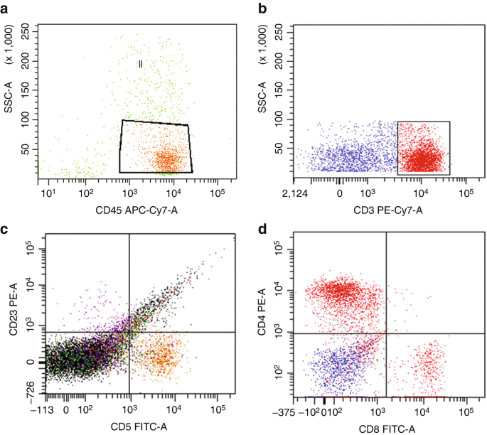
Flow Cytometry Of Mature And Immature T Cell Lymphoma Springerlink

Hodgkin S Disease Ask Hematologist Understand Hematology Non Hodgkins Lymphoma Hodgkins Lymphoma Lymphoma Cancer